


The valgus deformation of the feet is most common. In some cases, however, traumatic lesions can occur with paralysis in the mature lifespan. The main symptoms of pathology are pain in the area of the feet and muscles of the lower leg, a visible violation of the feet and a change of gear. The disease is diagnosed with a clinical examination, x rays, electromyography etc. The treatment includes conservative and surgical methods. However, the appropriate effectiveness is only observed in reconstructive operations.
What is this disease?
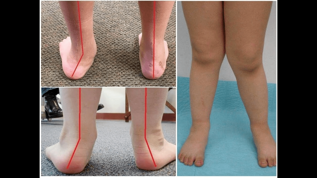
The valgus deformation is the curvature of the foot, which is characterized by the flattening of its longitudinal arch. Typically, the inner edge of the foot is lowered ("drop") and the heel turns out.
Due to his location, the foot of a person takes on the pressure of the entire mass of the human body. For this reason, it has a special anatomical structure that enables depreciation, the balance and stabilization of movements. However, an important component for the implementation of these tasks is the right stop -pound.
The most important problem of traumatology and orthopedics is the valgus deformation of the foot today. The meeting is estimated at 30-58%, with 2/3 of cases of innate disorders.
Pathology is largely socially significant, since all age groups of the population covers and also help to develop the spine, the early development of osteochondrosis and the arthrosis of the joints of the lower extremities.
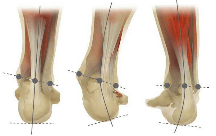
If you take your feet (if you look at it from behind), an X-like deformation is formed at ankle level: the ankles are in contact, while the heels are apart at a distance of 5 to 6 centimeters.
Most of the time, the pathology is naturally innate and is diagnosed in children in the hospital (or immediately after starting). A similar condition is adjusted for up to 5 years. After that, a child (in the absence of proper treatment) develops flat feet.
Why does it arise?
It is believed that the main reason for the occurrence of the valgus deformation of the foot is an inadequate function of the rear tibial muscle or the weakness of the band apparatus.
Today, other factors in the development of pathology are differentiated:
- Congenital anomalies with the wrong position of the feet bones or shortening of the tendons (vertical rambones, short piles);
- High -up disorders if the deformation of the feet spends the curvature of the spine;
- Traumatic lesions (fractures of the feet bones, lower legs, hips or knees, slide the band and tendon apparatus);
- Paralysis (immobilization) due to damage to the nervous system by encephalitis, poliomyelitis, stroke, violations of cerebrospinal melt for hernia, etc. ;
- Cramp (constant contraction) of the muscles of the lowerbone;
- Certified diseases: pathology of the bone system with vitamin D (rachitis), diabetes mellitus, osteoporosis (reduction in bone density), impaired thyroid and parathyroid glands, etc. ;
- Increased body weight, including rapid weight gain in menopause or during pregnancy.
The development of pathology is also facilitated by incorrectly selected shoes or excessive correction of the club foot in childhood.
Degree and stage of the disease
The severity of the pathology (power of the manifestation) is divided according to degree:
- Light with a vault of 1, 5 to 2 centimeters and an angle of inclination of the heel to 15 degrees;
- The average when the bow is flattened up to the 1st centimeter and the angle decreases to 10 degrees;
- Heavy at the height of the vault of up to 0, 5 cm and the angle of inclination of the heel is 5 degrees.
Depending on the participation of certain structures, the following stages of curvature are differentiated:
- There are no bone deformations, pain is determined on the inner surface of the ankle (in the area of fastening the tibia muscle);
- The curvature is light, the heel is slightly rejected;
- The foot is allocated and the deformation is fixed (not correctly corrected);
- Wing is not only observed in the foot, but also in the ankle joint.
Symptoms
In the first stage, patients are disturbed by periodic pain after long walks or long vertical loads (standing or sitting on the foot). As a rule, pain syndrome increases when walking in not properly selected shoes. The next stage of the disease is connected to the occurrence of the curvature of the foot: the patients in the standing position do not rely on the outside edge of the foot, but on their entire area. A slight change in the passage is observed.
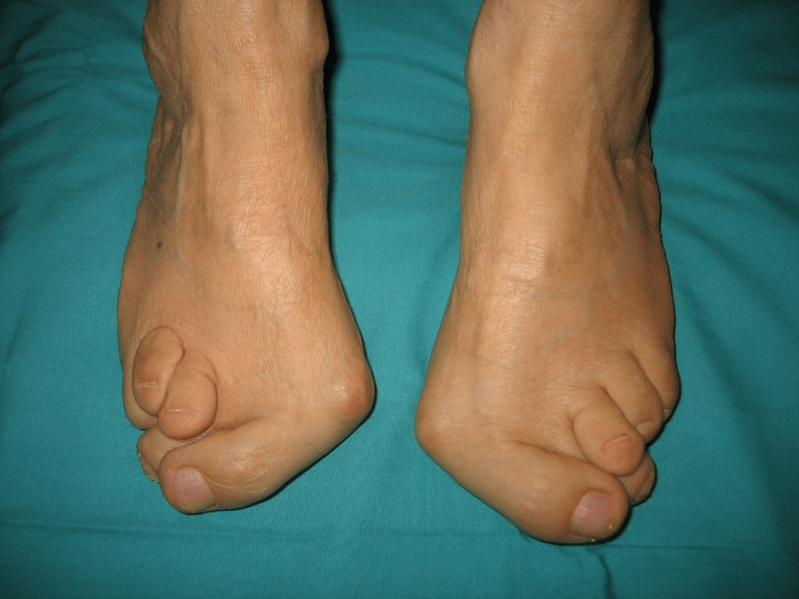
In the third stage, the lead of the thawed bone is determined (noticeably lower than the ankle on the inner surface of the ankle) and a strong detour of the heel outdoors (the patient is on the inner edge of the heel bone). The advanced valgus deformation of the feet is characterized by a pronounced curvature of the foot itself and ankle joint. The patients complain about severe pain in the lower leg muscles and a significant gang injury: the knees rub against each other, while the right and left foot is at a distance.
The severe curvature of the feet is often deformed by deformation of the spine (scoliosis with different positions of the shoulders and wings of the pool), osteochondrosis (damage to the intervertebral disc with the formation of a hernia and hip rod), osteochondrosis (damage to the Schwandebal disc with the formation of a hernia and hip -rod).
How do I diagnose?
The diagnosis of the curvature of the foot consists of:
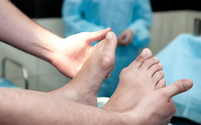
- The clinical inspection, in which the orthopedist recognizes a decrease in the floors, the deviation of the heel and rambones, the visible "disappear" of the outside and the lead of the inner ankles.
- X -Ray - an affordable and informative method with which you can determine the change in the angle of the bone incline and the linear parameters of your relationship. These indicators are necessary to make a final diagnosis and to clarify the degree of deformation.
- The method for registering steps to determine the exact state of function of the limb. The method is to register the time of supporting individual foot parts when carrying out a step. In the course of the study, the phases of the foot roller are also examined, which reflect the balance of the muscles of the lower extremity.
- Dynamic electromyography, which records the electrical activity of the muscles examined and their dependence on the step phase.
- Photoplesmography with digital processing, with which you can get all standard indicators and determine the type/degree of curvature with high accuracy.
An additional consultation of a neurologist (with deformations due to spasms or paralysis), the endocrinologist (in the case of diabetes or disorders of the thyroid/parathydent gland) and gynecologist (if the threat) may be necessary. If the curvature of the foot occurred against the background of osteoporosis, the densitometry is necessary - the examination of the bone density.
Treatment
A distinction is made between the main methods for the treatment of the valgus curvature of the feet. Do not destroy pain with ointments and injections!
Conservative approach
However, this type of help aims to get rid of the symptoms of the disease does not remove the basic cause of the pathology.
The technology includes:
- the use of orthopedic insoles to support the I Plus bone, the arch and the elimination of the valgus lesions of the middle and foot base;
- Taping - lead foot and ankles with special adhesive tapes with correct elasticity. The adhesive tape is worn around the clock within 3 to 5 days, according to which they are replaced;
- sew orthopedic shoes according to individual standards;
- The use of orthosis and other fixing devices on the foot and on the ankle.
Conservative methods also include physiotherapy methods (ozokerites, paraffin applications, electrophoresis, magnetic effects), massage and a complex of physiotherapy exercises that were developed for a specific clinical case. Be careful! Nowadays, most experts prefer surgical treatment methods because conservative therapy is ineffective (according to statistics, it is useless in 60% of cases).
Surgical intervention
The operating volume and its guy depend on the direct stage of the disease. Therefore, the first degree of valgus deformation is treated by synovectomy (removal of the tendon cover to correct the general voltage) or the osteotomy (dissection) of the heel in order to return to the anatomically correct position. In the second stage of the development of the disease, the transplantation of the tendons of the fingers is used. Such an intervention is usually carried out against the background of the dissection of the heel or a RAM-lifted arthrodesis (surgical immobilization of the joint between the RAM and Scapoid bones).
The curvature of the III degree requires an arthrodesis of several foot fuks at the same time: the plus-free five-cup cubic and RAM-derived. Such three points of immobilization are often supplemented by the dissection of the heel bone. In the IV pathology, reconstructive operations are not only required on the foot, but also on the ankle. In this case, the instability of the band apparatus using transplantation (from your own body or artificial materials) is adapted. The operating volume on the foot itself is the same as with the III curvature degree.
Recreational period
The rehabilitation includes for 2 months without support on the operated leg. At the same time, the patient has to wear removable plaster -longshit from 1, 5 to 3 months. Active movements in the operated foot are recommended to start after 1, 5 months after the operation. On the 3rd month, a complex of strengthening physical education is introduced. Afterwards, however, patients are prohibited from bumpy walking and active sports activities. It is worth noting that it is possible to assess the end result of the operation only six months later.
Preventive measures
The prevention of the Valgus deformation of the stop includes the following measures:
- Early correction of congenital anomalies with the improper arrangement of the feet bones or the shortening of the tendon grains (vertically thawed bones, short heel);
- Correction of posture disorders (scoliosis, etc. );
- Inclusion of the traumatic lesions (fractures of the foot bones, lower leg, thigh or knee joint, break of the tendon and tendon apparatus);
- Correct rehabilitation after paralysis (immobilization) due to the damage to the nervous system by encephalitis, poliomyelitis, stroke, violation of cerebrospinal roots for hernia, etc.
- Facilitation of the cramp (constant reduction) of the muscles of the lower leg;
- Therapy for simultaneous diseases: pathologies of the bone system with vitamin D (rachitis), diabetes mellitus, osteoporosis (reduction in bone density), impaired function of thyroid and parathyroid glands, etc. ;
- A decrease in body weight to normal (especially with a rapid weight gain in postmenopause or due to pregnancy);
- Selection of orthopedic shoes or the use of supinators;
- Moderate correction of club foot without "hyper -correction" improper treatment, which leads to a secondary valgus excretion of the feet.
Preventing the progression of the disease is to use conservative methods and early reconstructive operations. In this case, physical activity is limited to prevent the destruction and curvature of the ankles. Remember that the timely treatment of the valgus deformation of the feet not only improves the quality of life of patients, but also the development of osteochondrosis and osteoarthritis of the knee or hip joints!